近年来,癌症研究人员越来越关注细胞外基质 (ECM) 及其在癌症生长和扩散中的作用。当前的研究趋势逐渐从单纯针对癌细胞,转向同时考虑其周围的环境,尤其是 ECM 中发生的变化。
ECM 是由蛋白质和其他分子组成的网络结构,为组织提供结构支撑,并帮助细胞有序排列。但它不仅仅是一个物理框架,还参与调控多种关键的细胞过程,如生长、迁移、生存和分化。
在肿瘤微环境 (TME) 中,ECM 发生显著变化。其重塑过程有时会导致免疫环境减弱,从而使癌细胞免受自然免疫应答的攻击。这些变化还可能为肿瘤的生长与扩散创造有利条件,并通过阻断药物输送、促进治疗耐受性,使癌症更难治疗。随着我们对 ECM 的了解不断加深,它正日益被视为一种治疗靶标。研究人员正在探索如何靶向 ECM 的特定组成部分,以改变其行为,使其不再为肿瘤提供保护,而是转而抑制肿瘤的发展。
/26836_IHC%20analysis%20using%20Fibronectin%20antibody_thumbnail.jpeg?width=117&height=153&name=26836_IHC%20analysis%20using%20Fibronectin%20antibody_thumbnail.jpeg)
在这里,我们将介绍一些被作为潜在靶标研究的关键 ECM 标记物,包括支持肿瘤生长的结构蛋白、与肿瘤进展相关的信号蛋白以及重塑 TME 的 ECM 修饰蛋白。
<Jump to the product list at the end of this post>
结构蛋白:ECM 的基础
结构蛋白构成了 ECM 的骨架。它们负责组织的结构构建和稳定维持——但在癌症中,这些蛋白却可能会促进肿瘤的生长,并增强其对治疗的耐受性。
- 胶原蛋白约占 ECM 的 90%。1 虽然胶原蛋白对于健康组织至关重要,但肿瘤中胶原蛋白含量过高常与预后不良相关。胶原蛋白不仅可以促进癌细胞的生长和扩散,还能增强其对治疗的耐受性。2 胶原蛋白的关键标记物包括 COL1A1、COL11A1 和 COL6A。
/66948_IHC%20analysis%20using%20COL1A1%20monoclonal%20antibody_small.png?width=507&height=316&name=66948_IHC%20analysis%20using%20COL1A1%20monoclonal%20antibody_small.png)
使用单克隆抗体 COL1A1 (E3E1X) Mouse mAb #66948 对石蜡包埋的人类非小细胞肺癌组织进行免疫组织化学 (IHC) 分析。
- 层粘连蛋白是细胞基底膜结构的重要组成部分。1 它们使癌细胞能够附着于基底膜表面,进而在体内迁移并侵袭其他组织,在癌症转移过程中发挥关键作用。3 层粘连蛋白由三种不同的多肽链组成:α (LAMA)、β (LAMB) 和 γ (LAMC)。
/53884_LAMC2_ECM%20TME%20marker_A-431%20cells.png?width=507&height=252&name=53884_LAMC2_ECM%20TME%20marker_A-431%20cells.png)
使用重组单克隆抗体 LAMC2 (E9F7M) Rabbit mAb #53884(绿色)、DyLight™ 554 Phalloidin #13054(红色)和 DAPI #4083(蓝色)对 A-431 细胞(表皮样癌的模型人类细胞系)进行免疫荧光分析。
- 弹性蛋白通常为组织提供柔韧性和弹性。然而,当其发生降解时,会产生弹性蛋白衍生肽 (EDP),而研究表明这些肽可促进肿瘤发展。1,4
促肿瘤蛋白:促进生长和扩散
一些 ECM 蛋白不仅可以支持结构,还能主动促进肿瘤进展。接下来的一组标记物与肿瘤侵袭性、转移性增强以及治疗耐受性增加密切相关。
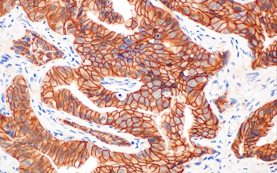
- 纤连蛋白已被证明有助于癌细胞生长和扩散。在上皮-间质转化 (EMT) 过程中,其表达水平上调,并与肿瘤侵袭性和转移能力的增强密切相关。因此,高水平的纤连蛋白可能表明癌细胞已向间充质状态转变。
/26836_IHC%20analysis%20using%20Fibronectin%20antibody%20in%20breast%20cancer.png?width=490&height=304&name=26836_IHC%20analysis%20using%20Fibronectin%20antibody%20in%20breast%20cancer.png)
使用重组单克隆抗体 Fibronectin/FN1 (E5H6X) Rabbit mAb #26836 对石蜡包埋的人乳腺导管癌组织进行免疫组织化学分析。
- Tenascin-C 可促进 TME 内的细胞迁移,一些研究表明它对于肿瘤细胞的转移至关重要。5 这种肿瘤基质成分在高度侵袭性癌症中通常呈高水平表达。
- Thrombospondin-1 是一种调节细胞粘附、迁移、细胞凋亡、炎症、血管功能和癌症发展的 ECM 蛋白。它还可以通过阻止新血管形成(血管生成)来减缓肿瘤的生长。6
/37879_WB%20analysis%20thrombospodin-1%20antibody.jpeg?width=520&height=350&name=37879_WB%20analysis%20thrombospodin-1%20antibody.jpeg)
使用 Thrombospondin-1 (D7E5F) Rabbit mAb #37879 对不同上皮细胞、上皮样细胞和成纤维细胞系(ACHN、ScaBER、LN18、mIMCD-3 和 MEF)提取物进行蛋白质印迹分析。
- 骨桥蛋白 (OPN) 已被证明参与细胞黏附和迁移,从而进一步促进肿瘤的转移。它还在肿瘤发生过程中发挥作用,可促使正常细胞转化为癌细胞,进而加速疾病进展。7 目前,人们正在将其作为潜在的治疗靶标进行研究。8
/88742_IHC%20analysis%20using%20osteopntin%20SPP1%20antibody.jpg?width=501&height=311&name=88742_IHC%20analysis%20using%20osteopntin%20SPP1%20antibody.jpg)
使用重组单克隆抗体 Osteopontin/SPP1 (E9Z1D) Rabbit mAb #88742 对石蜡包埋的小鼠卵巢组织进行免疫组织化学分析。
ECM 修饰蛋白:重塑肿瘤微环境
最后一组蛋白质,即 ECM 修饰蛋白质,能够改变 ECM,从而导致癌细胞扩散。
- 透明质酸是 ECM 中的一种糖胺聚糖 (GAG),可影响肿瘤细胞增殖、凋亡、血管生成和转移。它形成 TME 的屏障,阻碍药物输送到肿瘤部位。9
/53721_IHC%20analysis%20hyaluronan%20staining%20kit.jpg?width=500&height=336&name=53721_IHC%20analysis%20hyaluronan%20staining%20kit.jpg)
使用 Hyaluronan Complete Tissue Staining Kit (Alexa Fluor® 488) #53721(绿色)和 DAPI #4083(蓝色)对石蜡包埋的人结肠腺癌组织进行免疫组织化学分析。
- 赖氨酰氧化酶 (LOX) 是一种参与胶原蛋白和弹性蛋白之间交联形成的酶。它通过强化 ECM 来促进肿瘤生长。
- 基质金属蛋白酶 (MMP),特别是MMP-2、MMP-9、MMP-1 和 MMP-14(也称为 MT1-MMP),是分解 ECM 组分的酶,可使癌细胞更具扩散性。
/13667_WB%20analysis%20of%20MMP9%20antibody.jpeg?width=520&height=350&name=13667_WB%20analysis%20of%20MMP9%20antibody.jpeg)
使用重组单克隆抗体 MMP-9 (D6O3H) XP® Rabbit mAb #13667 对未经处理 (-) 或经过 TPA #4174(200 nM,48 小时;+)处理的 U-2 OS 细胞的提取物和浓缩培养基进行蛋白质印迹分析。正如预期的那样,MMP-9 是由 TPA 处理诱导的。
- Versican 是一种硫酸软骨素蛋白聚糖 (CSPG),可与 ECM 成分相互作用,从而促进癌细胞增殖、存活、血管生成、侵袭和转移。
- Periostin 是一种参与损伤后 ECM 重塑的基质细胞蛋白,可促进细胞增殖、存活和黏附,并最终导致癌细胞转移。
为什么细胞外基质在癌症研究中如此重要
随着该领域的不断发展,这些标记物有望在引领新一代癌症治疗中发挥核心作用。
研究人员通过深入揭示这些蛋白的独特性质及其对 ECM 在癌症进展中的影响,期望开辟创新的治疗策略。
参考文献
- Huang J, Zhang L, Wan D, et al. Extracellular matrix and its therapeutic potential for cancer treatment. Signal Transduct Target Ther. 2021;6(1):153. Published 2021 Apr 23. doi:10.1038/s41392-021-00544-0
- Fang M, Yuan J, Peng C, Li Y. Collagen as a double-edged sword in tumor progression. Tumour Biol. 2014;35(4):2871-2882. doi:10.1007/s13277-013-1511-7
- Aleman JD, Young CD, Karam SD, Wang XJ. Revisiting laminin and extracellular matrix remodeling in metastatic squamous cell carcinoma: What have we learned after more than four decades of research?. Mol Carcinog. 2023;62(1):5-23. doi:10.1002/mc.23417
-
Wang Y, Song EC, Resnick MB. Elastin in the Tumor Microenvironment. Adv Exp Med Biol. 2020;1272:1-16. doi:10.1007/978-3-030-48457-6_1
- Sun Z, Schwenzer A, Rupp T, et al. Tenascin-C Promotes Tumor Cell Migration and Metastasis through Integrin α9β1-Mediated YAP Inhibition. Cancer Res. 2018;78(4):950-961. doi:10.1158/0008-5472.CAN-17-1597
- Ming-Ping Wu, Li-Wha Wu, Cheng-Yang Chou (2016) The anticancer potential of thrombospondin-1 by inhibiting angiogenesis and stroma reaction during cervical carcinogenesis, Gynecology and Minimally Invasive Therapy, Volume 5: 15, 2-8.
- Zhao H, Chen Q, Alam A, et al. The role of osteopontin in the progression of solid organ tumor. Cell Death Dis. 2018;9(3):356. Published 2018 Mar 2. doi:10.1038/s41419-018-0391-6
- Bandopadhyay M, Bulbule A, Butti R, et al. Osteopontin as a therapeutic target for cancer. Expert Opin Ther Targets. 2014;18(8):883-895. doi:10.1517/14728222.2014.925447
- Zhao J, Chen J, Li C, Xiang H, Miao X. Hyaluronidase overcomes the extracellular matrix barrier to enhance local drug delivery. Eur J Pharm Biopharm. 2024;203:114474. doi:10.1016/j.ejpb.2024.114474


 沪公网安备31011502018823号